
Neuroimaging Pearls- A case of cervical internal carotid artery (ICA) dissection
A case of cervical ICA dissection
Abstract
A 58-year-old male with no underlying comorbidities presented with sudden onset weakness of the right side of the body with a preceding history of neck pain.
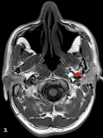
Magnetic resonance imaging (MRI) with Time of flight MR angiography (TOF-MRA) was performed. Axial diffusion weighted trace images (Fig. 1 and 2) show patchy areas of diffusion restriction in the left middle cerebral artery (MCA) territory. Axial T1 weighted imaged (Fig. 3) shows luminal expansion with eccentric T1 hyperintensity involving the left distal cervical internal carotid artery (red arrow) suggestive of intramural hematoma with a small residual lumen (white arrowhead). Sagittal reformat of TOF-MRA (Fig. 4) shows a linear, spiral hypointensity within the distal left cervical ICA (white arrow) suggestive of a dissection flap with hyperintense false lumen seen anteriorly (black asterisk). Note the expanded profile of the vessel. These imaging findings are characteristic of a dissection involving the distal cervical ICA. The intracranial vasculature was patent. The patient was managed conservatively on anticoagulants.
Downloads

This work is licensed under a Creative Commons Attribution 4.0 International License.


